CHAPTER - 18 : BODY FLUIDS AND CIRCULATION

All parts
of the body require nourishment and oxygen, and metabolic wastes need to
be removed from the body. So there is a need to transport various
substances like digested food materials to provide energy and growth of
the body, hormones, metabolic wastes, enzymes, various gases (oxygen and
carbon dioxide) etc. from one part of the body to other. These
functions are carried out by an extracellular fluid, which flows
throughout the body. This flow is known as circulation and this
transport of substances is done by a system is called circulatory
system.
Functions of the circulatory system :
· It transports nutrients from their sites of absorption to different tissues and organs for storage, oxidation or synthesis of tissue components.
· It also carries waste products of metabolism from different tissues to the organs meant for their excretion from the body.
· It transports respiratory gases between the respiratory organs and the tissues.
· It carries metabolic intermediates from one tissue to another for their further metabolism; for example, blood carries lactic acid from muscles to the liver for its oxidation.
· It also transports informational molecules such as hormones, from their sites of origin to the tissues.
· It uniformly distributes water, H+, chemical substances to all over the body.
Blood Vascular System :
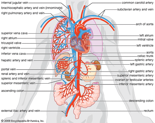
Higher animals have a well-developed circulatory system so that transport of substances in the body can be done very effectively. In them, the circulatory system consists of a central pumping organ called as heart and various blood vessels (arteries, veins and capillaries). Arteries conduct the blood from the heart to other tissues; veins bring blood from other tissues to the heart. Some of the invertebrates and all vertebrates possess this system. The circulatory system was first discovered and demonstrated by William Harvey.
The blood vascular system may be of two types, the open and the closed circulatory systems.
Open circulatory system :
In many advanced invertebrates such as prawns, insects and molluscs, the blood does not remain confined to blood vessels but it flows freely through the body cavity and channels called lacunae and sinuses in the tissues. The body cavity is known as hemocoele and the blood is hemolymph. In insects, the tissues are in direct contact with the blood. Hemolymph circulates in the whole body due to the contractile activity of heart.
Closed circulatory system :
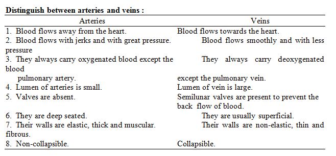
In closed circulatory system the blood flows through proper blood vessels named arteries, veins and blood capillaries. Arteries within the tissues divide into arterioles, which then branch further to form capillaries. Capillaries then unite to form venules, which come out of the tissues and veins. Arteries have thick, elastic and muscular walls which are made up of three concentric layers viz., tunica externa, tunica media and tunica interna. All these layers have got smooth or involuntary muscles. Contraction and relaxation of smooth muscles alter the diameter of arteries and thus regulate the flow of blood through them. Capillaries are extremely fine, thin blood vessels the walls of which are made of a single layer of endothelial cells. The muscles and elastic fibers are absent in them. These capillaries are highly permeable to water and small macromolecules. Various nutrients, respiratory gases, metabolites and other substances are exchanged between the blood and tissues through these capillaries.
Structurally veins resemble arteries except that the three layers are very thin and more elastic. In the veins the muscles and elastic connective tissues are poorly developed. But the collagen fibers of the outer layer are very well developed. In most of the veins the middle coat is extremely thin with practically no muscles. In many veins semilunar valves are present in their lumen. These valves allow the flow of the blood only in one direction i.e., towards the heart.
The heart :· It transports nutrients from their sites of absorption to different tissues and organs for storage, oxidation or synthesis of tissue components.
· It also carries waste products of metabolism from different tissues to the organs meant for their excretion from the body.
· It transports respiratory gases between the respiratory organs and the tissues.
· It carries metabolic intermediates from one tissue to another for their further metabolism; for example, blood carries lactic acid from muscles to the liver for its oxidation.
· It also transports informational molecules such as hormones, from their sites of origin to the tissues.
· It uniformly distributes water, H+, chemical substances to all over the body.
Blood Vascular System :
Higher animals have a well-developed circulatory system so that transport of substances in the body can be done very effectively. In them, the circulatory system consists of a central pumping organ called as heart and various blood vessels (arteries, veins and capillaries). Arteries conduct the blood from the heart to other tissues; veins bring blood from other tissues to the heart. Some of the invertebrates and all vertebrates possess this system. The circulatory system was first discovered and demonstrated by William Harvey.
The blood vascular system may be of two types, the open and the closed circulatory systems.
Open circulatory system :
In many advanced invertebrates such as prawns, insects and molluscs, the blood does not remain confined to blood vessels but it flows freely through the body cavity and channels called lacunae and sinuses in the tissues. The body cavity is known as hemocoele and the blood is hemolymph. In insects, the tissues are in direct contact with the blood. Hemolymph circulates in the whole body due to the contractile activity of heart.
Closed circulatory system :
In closed circulatory system the blood flows through proper blood vessels named arteries, veins and blood capillaries. Arteries within the tissues divide into arterioles, which then branch further to form capillaries. Capillaries then unite to form venules, which come out of the tissues and veins. Arteries have thick, elastic and muscular walls which are made up of three concentric layers viz., tunica externa, tunica media and tunica interna. All these layers have got smooth or involuntary muscles. Contraction and relaxation of smooth muscles alter the diameter of arteries and thus regulate the flow of blood through them. Capillaries are extremely fine, thin blood vessels the walls of which are made of a single layer of endothelial cells. The muscles and elastic fibers are absent in them. These capillaries are highly permeable to water and small macromolecules. Various nutrients, respiratory gases, metabolites and other substances are exchanged between the blood and tissues through these capillaries.
Structurally veins resemble arteries except that the three layers are very thin and more elastic. In the veins the muscles and elastic connective tissues are poorly developed. But the collagen fibers of the outer layer are very well developed. In most of the veins the middle coat is extremely thin with practically no muscles. In many veins semilunar valves are present in their lumen. These valves allow the flow of the blood only in one direction i.e., towards the heart.
The heart is the central pumping organ of the blood vascular system. It is a hollow muscular structure and is made up of cardiac muscles. It works throughout life rhythmically without getting tired. It is enclosed in a double membraneous sac called pericardium that is filled with pericardial fluid. Mainly there are two chambers in a heart – auricle or atrium that receives the deoxygenated blood from various parts of the body; and a ventricle that distributes the oxygenated blood to the body. The number of these chambers varies in different animals.
In fishes, the heart is only two chambered – one auricle and one ventricle. Both these chambers contain deoxygenated blood.
In amphibians, the auricle is divided into right and left auricles. The blood after oxygenation from lungs is returned back to left auricle. Right auricle receives deoxygenated blood from various parts of the body. However, in the ventricle there is mixing up of deoxygenated and oxygenated blood.
In reptiles (except crocodiles), the division of the ventricle also starts but it is not complete. So the heart is incompletely four-chambered. However, there are two auricles- left and right auricles. In them the oxygenated and deoxygenated blood are kept separate. But in the ventricle, this separation is not perfect.
Crocodiles, birds and mammals have a complete four-chambered heart. In them the ventricle septum is complete so that there is no mixing up of oxygenated and deoxygenated blood at all.
A structure called sinus venosus is present in the hearts of fishes, amphibians and reptiles. It receives deoxygenated blood from anterior and posterior caval veins and then that blood is poured into the heart. There is no sinus venosus in mammals.
Human Heart :
The mammalian heart including man is a hollow, cone-shaped, muscular structure that lies in the thoracic cavity above the diaphragm and in between the two lungs. It is about the size of a fist measuring about 12 cm in length and 9 cm in breadth. It weighs about 300 grams. It is a four chambered organ-two atria or auricles and two ventricles. Deoxygenated blood is received into right auricle by superior vena cava (from anterior region) and inferior vena cava (from posterior region) of the body. These vena cavae open directly into right auricle as there is no sinus venosus. Right auricle also gets blood from coronary veins (from the heart muscles itself). The right and left auricles are separated by interauricular septum. Similarly, right and left ventricles are also separated by interventricular septum. Deoxygenated blood is then passed from the right auricle to the right ventricle through the atrioventricular aperture guarded by tricuspid valve (having three flaps). The blood is then pumped into lungs for oxygenation via pulmonary artery. After oxygenation, the blood is brought back into left auricle via four pulmonary veins. From left auricle, blood (now oxygenated) goes to left ventricle through atrio-ventricular aperture and this opening is regulated by bicuspid (having two flaps) or mitral valve. The left ventricle has also got chordae tendinae and papillary muscles which prevent the valves (both bicuspid and tricuspid) from being pushed into auricles at the time of ventricular contraction. Thus the walls of left ventricle are thicker than the walls of right ventricle. The oxygenated blood from left ventricle is then distributed to all parts of the body with the help of aorta. The openings of the aorta and other major arteries are guarded by semilunar valves that prevent the back flow of blood.
Course of Circulation through Mammalian Heart :
During a heart beat, there is contraction and relaxation of auricles and ventricles in a specific sequence. The contraction phase is known as systole, while relaxation phase is known as diastole. Various series of events that occur during a heart beat is known as cardiac cycle.
When both the auricles and ventricles are in relaxed or diastolic phase. This is referred to as joint diastole. During this phase, the blood flows into the auricles from the superior vena cava and inferior vena cava. The blood also flows from the auricles to their respective ventricles through the atrio-ventricular valve. There is no flow of blood from the ventricles to the aorta and its main arteries as the semilunar valves remain closed in this phase.
At the end of joint diastole, the next heart beat starts with the contraction of atria (atrial systole). In this phase, it now forces most of its blood into the ventricle, which is still in the diastolic phase. During auricular systole, the blood cannot pass back into the superior and inferior vena cava because they are compressed by the auricular contraction and their openings to the auricles are blocked. Thus auricles act as main vessel to collect and pump the venous blood into the ventricles. Thus at the end of auricular systole, the auricles get empty.
After the atrial systole is over, the auricular muscles relax and it enters into auricular diastolic phase. During auricular diastole, it again gets filled up with the venous blood coming from the superior and inferior vena cavae. Along with the auricular diastole, the ventricular systole starts. This results in an increased pressure of blood in the ventricle and it rises more than the pressure of blood in the auricle. Soon the atrio-ventricular valves are closed and thus the back flow of blood is prevented. This closure of AV-valve at the beginning of ventricular systole produces a sound “lubb” and is known as the first heart sound. Initially, when the ventricle starts contracting, the pressure of blood within it is lower than the pressure of blood within the aorta and so the semilunar valves do not open. Therefore, the ventricle contracts as a closed chamber. As the ventricular systole progresses more, the pressure of blood within the ventricle increases more than that of aorta as a result the semilunar valves now open and blood flows (with a speed) into the aorta and its main branches. The back flow of blood in the auricles is prevented, as the AV-valves remain closed.
Now at the end of ventricular systole, ventricular diastole starts. As the auricles are still continuing with their diastole, so all the four chambers are now in diastole. This is known as joint diastole. In the ventricular diastolic phase, the pressure of blood in the ventricles falls below the pressure of blood in the aorta, so the semilunar valves get closed to prevent the back flow of blood from the aorta to the ventricles. This closure of semilunar valves at the beginning of ventricular diastole produces a sound “dup” and is known as the second heart sound. After the closure of the semilunar valves, the ventricles become closed chambers again. Also, as the ventricular pressure is more than the atrial pressure, so the AV-valves remain closed. However, as the ventricular diastole continues, the pressure of blood in the ventricles falls below the pressure of blood in the auricles. At this point, the AV-valves open and blood starts flowing again from the relaxed auricles to the relaxed ventricles. Now when the joint diastole is over, the auricular systole starts and the blood is pumped into the ventricles.
Heart Rate and Pulse:
In the resting condition, human heart beats at the rate of about 70 times per minute. But, the heart beat rate increases during exercise, fever, and emotions like anger and fear. During each heart beat, the blood is pumped from the ventricles of the heart into the aorta to be distributed to all parts of the body. This happens during the ventricular systole and is repeated every 0.8 seconds. The blood from aorta then goes to other arteries of the parts. This causes a rhythmic contraction in the aorta and its main arteries. It can be felt as regular jerks or pulse in the regions where arteries are present superficially like wrist , neck and temples. This is known as arterial pulse.
The pulse rate is, therefore, same as that of heart beat rate. This heart beat rate differs from species to species. In general, the smaller the animal, the greater the heart beat. Hence, larger animals have lower heart rates. For example, an elephant has a normal heart beat rate of about 25 times per minute, where as mouse has a normal heart beat rate of several hundreds per minutes.
Automatic rhythmicity of the heart:
The mammalian heart is a myogenic heart i.e., the heart beat originate from a muscle (but it is regulated by nerves). In the right atrium near the region where superior vena cava opens, a specialised muscle called sinu-auricular node (SA-node) is present from where the heart beat originates. It is also called as pace maker and is richly supplied with blood capillaries. A wave of contraction (systole) originates from it and spreads over to the whole heart.
At the junction of right atrium and right ventricle, a tissue called auriculo-ventricular node (AV-node) is present that picks up the wave of contraction propagated by SA-node. This is also known as bundle of His. Branches of this spread over the ventricle forming the Purkinje system. The wave of contraction spreads over the ventricle through AV-node and its Purkinje system.
The heart is supplied with vagus (parasympathetic) and sympathetic nerve fibers. The vagus nerve is inhibitory and so when stimulated slows down the heart beat; while the sympathetic nerve is acceleratory and so when stimulated fastens the heart beat. This happens because these nerves release chemicals (hormones) when stimulated.
Circulation:
In vertebrates, the heart pumps blood into a closed circulatory system. The left ventricle ejects blood into the aorta, which gives off arteries to tissues and organs(except lungs), then the blood is returned from these tissues and organs through two veins, superior and inferior vanae cavae to the right atrium. This is known as the systemic circulation. The right ventricle pumps blood into the pulmonary trunk which divides into pulmonary arteries going to the lungs; then blood is returned to the left atrium from the lungs through the pulmonary veins. This is called the pulmonary circulation.
In some cases, before the blood can finally return to the heart, a vein returning blood from a system of capillaries divides again into a second capillary system in the tissues. Such type of vein is called as portal vein; and it constitutes a portal system along with the capillary system to which it supplies blood.
Veins after collecting deoxygenated blood from the organs normally pour the blood into right auricle. But sometimes, they pour their blood into some other organ by the portal veins before the heart. The blood from that organ is then collected and poured into the heart. For example, a hepatic portal vein returns blood from the intestine and breaks into a portal system of capillaries in the liver. This helps the absorbed nutrients from the small intestine to reach first into the liver via the hepatic portal vein. The cells of the liver can take up these nutrients. Similarly, the blood coming from hypothalamus may be poured into anterior pituitary by a hypophysial portal vein. This portal system enables the hormones of hypothalamus to reach the anterior pituitary.
Arterial Blood Pressure:
The pumping action of the heart maintains a pressure of blood in the arteries. This is called Arterial blood pressure. It helps to pump blood at a high velocity along the arteries in the closed circulatory system. The blood pressure is far lower in the open circulatory system.
Blood Flow in Veins:
The blood pressure is low in veins, because the blood flows through narrow arterioles and capillaries to enter wider veins. At many places in the body, this blood pressure is not sufficient to drive the blood through the veins back to the heart. Veins have thinner walls than arteries and are more easily compressed. There are also many valves inside the veins. These valves permit the flow of blood in the veins towards the heart and prevent blood flow in the reverse direction. Contraction of muscles and changes of body posture compresses the veins to move the blood inside them. During this both cases, blood moves towards the heart only, because the venous valves prevent the blood flow in the opposite direction. This is a major process for venous blood flow.
For example, if a person stands immobile for a long time, blood flow in the leg veins remains suspended. This may lead to an accumulation of fluid in his leg tissues and a consequent swelling of his feet. If he walks for some time, the swelling subsides as blood begins to circulate again in the veins.
Lymph and Tissue Fluid:
It occurs in the spaces in between the cells of a tissue and is called as interstitial fluid or tissue fluid. The exchange of any material (solid, liquid or a gas) that occurs between the blood and the tissue cells always takes place through this fluid. Under the pressure of blood in the capillaries some of the water and desired solutes are filtered out from the blood plasma into the tissue spaces to form the tissue fluid. The composition of this tissue fluid is very similar to that of plasma except that it has much less protein. Proteins are less because some of the proteins are not filtered out from the capillary walls (impermeable).
Some of the tissue fluid enters tiny channels called lymph vessels and the fluid collected in them is called lymph and this system is known as lymphatic system. These lymph vessels unite to form larger lymph vessels which ultimately drain into two large lymph vessels called thoracic duct and the right lymphatic duct. These open into veins returning the lymph finally into venous blood and so in the general circulatory system. This movement of lymph is mainly due to the squeezing action of the surrounding muscles. So the lymphatic system is slow and uncertain. Exercise increases the rate of lymph circulation.
Generally, the rate of lymph formation is equal to the rate of its return to the blood stream. But sometimes, the formation rate of lymph exceeds the rate of its return to blood. The increased volume of fluid around the cells then creates a swelling, called dropsy or oedema.
Functions of Lymph:
1. It serves to return interstitial fluid into blood.
2. The plasma proteins macromolecules synthesized by the liver cells, cannot pass into the blood vessels, but can diffuse into the lymph vessels through their wall and they come to the blood through lymph.
3. It also carries absorbed fats and lipids from the small intestine to the blood in the form of chylomicron droplets.
Disorders of Circulatory System:
1. High Blood Pressure (Hypertension): It is the term for blood pressurethat is higher than normal of 120/80. In the instrument 120 is Systolic / pumping pressure, 80 is Diastolic, resting pressure. If repeated check which shows 140/90 and higher shows Hypertension. It may leads to heart diseases and also affect vital organs like brain and kidney.
2. Coronary Artery Disease (CAD) : it is also known as Atherosclerosis, due to damage in the blood vessels of heart tissues. Basically it it due to, deposition excess of Calcium, fat, cholesterol and fibrous tissues which makes the lumen of arteries narrower.
3. Angina: It is also known as “angina pectoris”, it is symptom of acute chest pain due to less oxygen supply to heart.
4. Heart Failure: It is the state when heart is not pumping blood effectively to other organs of the body.
