CHAPTER - 22 : CHEMICAL COORDINATION AND INTEGRATION

INTRO
The glands are secretory organs and are of two main types, viz. (i) exocrine glands and (ii) endocrine or ductless glands. Endocrine glands secrete active substances called Hormones. Hormones are informational molecules. They are secreted in response to changes in the environment inside or outside the body. They are secreted into the blood, which distributes it all over the body, especially to their target organs and tissues.
Hormones are the chemical substances produced from an endocrine or a ductless gland. They may be defined as, “substances which are produced in one part of an organism and transferred to some other part where their physiological effects are observed”. Chemically they may be polypeptides, steroids and biogenic amines (non-protein compounds containing amino group).
Internal Environment of animal body is maintained in a steady state by
1. Autonomic Nervous System
2. Endocrine System
What are glands?
They are secretory organs.
Types of glands:
Exocrine glands – Duct glands - Enzymes
Endocrine glands – Ductless glands – Hormones
What are Hormones?
Chemical regulators / messengers / information molecules
Chemical nature of Hormones
Organic substances of varying complexity fall into two major classes;
1. Steroid Hormone
2. Amino acid Hormone
Characteristics of Hormone:
•Target organs / tissues
•Specific in their action
•Trace amount
Functions of Hormones
•Metabolic activities
•Homeostasis
•Morphogenic activities
•Mental activities
•Growth, maturation and regeneration
•Secondary sexual characters and reproductive activities
•Control of other endocrine glands
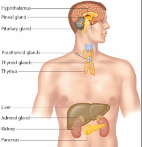
Endocrine glands in man
•Pituitary glands – In head
•Thyroid glands – In neck
•Parathyroid – embedded on thyroid gland
•Adrenals – upper end of kidney
•Thymus – on either side of trachea
•Gonads – In or below pelvic cavity
•Gastric – In the wall of stomach & intestine
•Pineal gland – dorsal side of brain
•The hypothalamus is a region of the brain that controls an immense number of bodily functions.
•The pituitary gland, also known as the hypophysis, is a roundish organ that lies immediately beneath
the hypothalamus
• It composed of two distinctive parts:
The anterior pituitary (adenohypophysis) & the posterior pituitary (neurohypophysis).
Characteristics of hormones:
· Hormones have more or less a specific role. The spectrum of action varies with the hormone. Some are highly selective, while others are more generalized.
· Hormones are produced from a tissue or an organ, and then they act on different tissues or organs i.e., they have a target organ to act or their activity is at a remote rate.
· Hormones can be easily transported via blood. They are poured into venous blood.
· They are active in minute concentrations, only a few picograms (10-12 g) or a few microgram (10-6 g). The number of hormone molecules per unit target tissue is definite.
· A hormone in its “primary action” affects one or a limited number of reactions and does not influence directly all other metabolic activities of the cell.
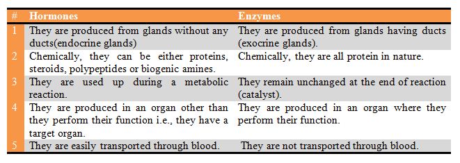
Differentiate hormones and enzymes:
Mammalian endocrine system:
The mammalian endocrine system consists of the following organs and tissues : Hypothalamus pituitary, thyroid, four parathyroids, two adrenals, two testes (male) or two ovaries (female), thymus, pineal, islet tissue of pancreas, and hormonal tissues on gastrointestinal tract. The hypothalamus is a nervous system of the brain, which is also integrated with the endocrine system and secretes hormones.
Hypothalamo-pituitary Axis:
Hypothalamus as an endocrine gland:
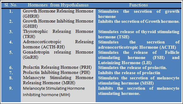
Hypothalamus is a part of the brain and consists of several masses of grey matter called hypothalamic nuclei. In fact, it forms the floor of the third cerebral ventricle of the brain. Neurons of the hypothalamic nuclei control the activity of pituitary gland. The hypothalamus is connected to the anterior lobe of pituitary by hypophyseal portal vessels. The hypothalamic nuclei or neurosecretory cells secrete several hormones called neurohormones that reach the anterior pituitary by hypophyseal portal vessels. These neurohormones control the secretions of the hormones from the anterior pituitary. These neurohormones are given below:
Pituitary as an endocrine gland :The mammalian endocrine system consists of the following organs and tissues : Hypothalamus pituitary, thyroid, four parathyroids, two adrenals, two testes (male) or two ovaries (female), thymus, pineal, islet tissue of pancreas, and hormonal tissues on gastrointestinal tract. The hypothalamus is a nervous system of the brain, which is also integrated with the endocrine system and secretes hormones.
Hypothalamo-pituitary Axis:
Hypothalamus as an endocrine gland:
Hypothalamus is a part of the brain and consists of several masses of grey matter called hypothalamic nuclei. In fact, it forms the floor of the third cerebral ventricle of the brain. Neurons of the hypothalamic nuclei control the activity of pituitary gland. The hypothalamus is connected to the anterior lobe of pituitary by hypophyseal portal vessels. The hypothalamic nuclei or neurosecretory cells secrete several hormones called neurohormones that reach the anterior pituitary by hypophyseal portal vessels. These neurohormones control the secretions of the hormones from the anterior pituitary. These neurohormones are given below:
Pituitary is a small body, about the size of a gram located on the ventral side of the diencephalon region of the brain. The pituitary hangs below the hypothalamus by a stalk called as infundibulum. The pituitary has three different parts viz. anterior lobe or adenohypophysis, intermediate lobe and posterior lobe or neurohypophysis. The adenohypophysis is compact and highly vascular. It is connected to the hypothalamus by hypophyseal portal vessels. The neurohypophysis is connected to the hypothalamus by nerve fibres. The anterior lobe of pituitary releases six hormones (all protein in nature) that control the activities of various other endocrine glands also. They are given below:
1. Growth hormone or Somatotrophic hormone (GH or STH). It is secreted by the Somatotrophic cells of anterior pituitary and regulates general body growth; increases the length of bones; control carbohydrate, protein, and fat metabolism; muscles and viscera growth; may counteract insulin to raise blood glucose levels etc. Its deficiency causes dwarfism in youngs, and acromicria (rarely) in adults – hypoactivity; while its excessive secretion – hyperactivity causes gigantism in youngs, and acromegaly in adults.
2. Adrenocortico-trophic hormone (ACTH). It is secreted by the Corticotrophic cells of anterior pituitary and controls the growth and secretion of adrenal cortex to release glucocorticoids – cortisol, cortisone etc. However, the secretion of mineralocorticoids by adrenal medulla is stimulated to a much less degree.
3. Thyroid stimulating hormone (TSH) or Thyrotrophic hormone or Thyrotropin. It is secreted by the Thyrotrophic cells of anterior pituitary and controls the growth and activity of the thyroid gland. It acts on thyroid to release its hormone – thyroxine.
4. Follicle stimulating hormone (FSH). It is also secreted by Gonadotrophic cells of anterior pituitary and increases the number and size (maturation) of graffian follicles in the ovaries in females; and stimulates spermatogenesis in males.
5. Luteinising hormone (LH) or interestitial cell stimulating hormone (ICSH). It is also secreted by Gonadotrophic cells. In females: (i) it completes the development of graffian follicles to its secretory stage and brings about ovulation along with FSH; (ii) it causes appearance, growth and maintenance of corpus luteum; (iii) it stimulates the secretion of progesterone from the ovaries. In males: it stimulates the development and functional activity of interestitial cells to produce testosterone.
6. Prolactin or Lactogenic hormone or Luteotrophic hormone (LTH). It helps in the growth of mammary glands during pregnancy and initiates the secretion of milk after child birth.
The posterior lobe of pituitary releases the following two peptide hormones. Both these hormones are synthesized in the hypothalamus and are carried to the posterior pituitary along with nerve fibres where they are stored. From posterior pituitary, they are released into the blood.
1. Vasopressin or Pitressin or Antidiuretic hormone (ADH). It is released from posterior pituitary in response to stress and dehydration. It increases the reabsorption of water in the distal convoluted tubules and the collecting tubules of kidney. So its deficiency in the body increases the urine flow causing diabetes insipidus. It also raises blood pressure by constricting the peripheral blood vessels.
Deficiency of ADH
Hypothalamic ("central") diabetes insipidus results from a deficiency in secretion of antidiuretic hormone from the posterior pituitary.
Nephrogenic diabetes insipidus occurs when the kidney is unable to respond to antidiuretic hormone
The major sign of either type of diabetes insipidus is excessive urine production.
2. Oxytocin or Pitocin. It is an important uterus-contracting hormone at the time of child birth. It also acts on mammary glands and helps in the expulsion of milk at the time of suckling. It is, therefore, also known as `milk-ejection hormone’ and `birth hormone’. It decreases the blood pressure by dilating the peripheral blood vessels (opposite to that of vasopressin).
Feed back inhibition of hormones:
Hypothalamus produces thyrotropin releasing factor (TRF) that acts on anterior pituitary to release thyroid stimulating hormone (TSH). This TSH then acts on thyroid to release its hormone – thyroxine. Now if the level of thyroxine in the blood is more, it will inhibit the hypothalamus to produce TRF, hence less of TSH and thyroxine. And if thyroxine is less, more of TRF is produced. Hypothalamus may also be inhibited or activated by the levels of TSH. This is known as feed back inhibition.
Thyroid Gland:
Thyroid hormones are derivatives of the amino acid tyrosine bound covalently to iodine.
The two principal thyroid hormones are: thyroxine (known affectionately as T4 or L-3,5, 3',5'-tetraiodothyronine) triiodotyronine (T3 or L-3,5,3'-triiodothyronine).
Thyroid epithelial cells - the cells responsible for synthesis of thyroid hormones – are arranged in spheres called thyroid follicles. Follicles are filled with colloid, a proteinaceous depot of thyroid hormone precursor.
Thyroid is found on the ventral side in the neck region of the body. At the base of larynx, it has two lateral lobes one on either side of trachea. Cells lining the thyroid follicles secrete two thyroid hormones, thyroxine and triiodothyronine. Both are iodinated forms of an amino-acid called thyronine and stored in the form of semifluid material (colloid) in the lumen of follicles. Whenever necessary, the hormones are released from the colloid to the blood.
Functions:
· Thyroid hormones increase the metabolic rate of the body, enhance heat production and maintain BMR.
· They also promote growth of body tissues-both physical growth and development of mental faculties are stimulated.
· They stimulate tissue differentiation; hence promote metamorphosis of tadpoles into adult frogs.
Disorders due to thyroid hormone imbalances:
· Excessive secretion of thyroid hormone (hyperthyroidism) results in exophthalmic goitre or Grave’s disease. It is accompanied by the bulging of the eye ball. It is associated with high metabolic rate, heart beat and blood pressure rises, restlessness, tremors, nervousness, rise in body temperature, etc.
· Less secretion of thyroid hormone (hypothyroidism) results in Myxedema in adult and cretinism (feeble mindedness) in children. The patient of myxedema suffers from low metabolic rate, slow heart rate, low body temperature and reproductive failure. Adminsitration of thyroid hormones cures the symptoms.
· For the synthesis of thyroid hormone (thyroxine), an important inorganic ion called iodine is needed in the body. Hence the dietary deficiency of iodine causes goitre in which thyroid gland enlarges in an effort to produce more thyroxine.
· Failure of thyroid hormone secretion in children slows body growth and mental development and reduces metabolic rate. The child becomes stunted and mentally retarded. The body temperature, heart rate and blood pressure are lower than normal. The patient is pot-bellied and pigeon chested and has a protruding tongue. This disease is known as cretinism.
Thyroid gland – Disorders
Hypothyroidism:
Reasons:
•Failure of thyroid gland
•Hyposecretion of TRH, TSH or Both
•Inadequate dietary iodine
Symptoms:
•Low metabolic activity
•Poor tolerance of cold
Hypothyroidism
Cretinism (in children):
Poor skeleton growth – dwarfism – mentally retarded – dry skin – thick tongue – lethargy – respiratory problems – constipation – neonatal jaundice.
Myxoedema (in adult):
Edema – facial tissues to swell and look fluffy.
Hyperthyroidism
Grave’s disease (exophthalmic goitre):
•An immune disease in which autoantibodies bind to and activate the thyroid-stimulating hormone receptor, leading to continual stimulation of thyroid hormone synthesis – edema behind eyes (exophthalmos) with – more often in females.
Parathyroids:
They are four pea-sized organs, clinging to the rear surface of thyroid, but are independent of thyroid structurally and functionally. This gland secretes parathormone (PTH) whose functions are as follows:
a) It regulates the calcium and phosphate balance between blood and other tissues.
b) It inhibits synthesis of collagen by osteoblasts and bone resorption by osteoclasts.
c) It helps in absorption of calcium from the intestine and reabsorption by kidneys.
Disorders:
a) Hypoparathyroidism:
It leads to deficiency of plasma calcium. Nerve and muscle action potentials rise leading to muscle twitches, spasm, etc., and the condition is called hypocalcemia or parathyroid tetany.
b) Hyperparathyroidism:
It causes demineralization of bones leading to their easy fracture and deformation. It may lead to osteitis fibrosa cystica.
Adrenals or Suprarenals:
There are two adrenal glands, one on the top of each kidney. Adrenal gland has an outer portion called adrenal cortex, and an inner portion called adrenal medulla. Both these parts differ in the nature of hormone produced and also with respect to their functions.
Adrenal cortex:
This part of adrenal gland is very important for the animal and its removal or destruction will kill the animal. It produces mainly three groups of steroid hormones viz., (i) glucocorticoids; (ii) mineralocorticoids; and (iii) sex corticoids.
(i) Glucocorticoids regulate the metabolism of carbohydrates, proteins and fats; they also increase the blood glucose level e.g., cortisone, cortisol and corticosterone. The secretion of glucocorticoid hormones is regulated by ACTH from anterior pituitary.
(ii) Mineralocorticoids are produced from the outermost cellular layer of the adrenal cortex. The main hormone in mammals and birds is aldosterone that reduces the sodium loss from the body in urine, sweat, saliva etc. by its active reabsorption from those fluids. It also increases the excretion of potassium, in an exchange with the absorption of sodium; retain water in the body along with sodium. Thus it regulates the ionic and water balance of the body. The secretion of aldosterone is stimulated by three factors : (a) a fall in the sodium ions in the plasma of blood; (b) a rise in the potassium ions in the plasma of blood ; (c) a fall in the volume of blood itself.
(iii) Gonadocorticoids: They stimulate the development of secondary sexual characters in males
Adrenal medulla:
It helps the body to prepare for stress or any emergency conditions. This part of adrenal, unlike cortex, is not so vital for the survival of the organism and so its removal will not cause death.
It produces two hormones called as adrenaline or epinephrine and nor-adrenaline or nor-epinephrine. Both these hormones act on tissues and organs that are supplied by sympathetic fibres and produce similar effects. Thus adrenal medulla and sympathetic system function as a closed integrated unit and is known as sympathetico-adrenal system.
Pancreas :
The pancreas comprises both exocrine and endocrine parts. The endocrine part of pancreas forms the islets of langerhans. They are small patches of cells present in the pancreas. They produce the following two hormones:
•The endocrine portion of the pancreas takes the form of many small clusters of cells called islets of Langerhans
•Pancreatic islets house three major cell types, each of which produces a different endocrine product:
•Alpha cells (A cells) secrete the hormone glucagon.
•Beta cells (B cells) produce insulin
•Delta cells (D cells) secrete the hormone somatostatin.
1. Insulin. It is protein hormone produced by Beta-cells of islets of langerhans. It regulates the amount of glucose in the blood by converting excess of glucose into glycogen (glycogenesis) which can be stored in the liver and muscles. Lack of insulin, therefore, results in excess glucose in blood (hyperglycemia) and it starts appearing in urine. This disease is known as diabetes mellitus.
Functions of Insulin:
· It increases the utilization of glucose in the tissues and facilitates the storage of glucose as glycogen in the liver and muscles. As a consequence, insulin lowers the blood glucose level in the body.
· It increases the synthesis of fats in the adipose tissues from fatty acids and also from glucose.
· It reduces the break down and oxidation of fats.
· It promotes protein synthesis from amino acids in the body tissues.
· It also reduces the breakdown of proteins in the body. Thus, insulin can be regarded as an anabolic hormone.
2. Glucagon. It is also protein hormone produced by Alpha-cells of islets of langerhans. It also regulates the amount of glucose in the blood by converting glycogen back to glucose whenever required. Thus its effect is opposite to that of insulin.
3. Somatostatin: It inhibits the secretion of glucagons and insulin.
Insulin Disorders
Hyperglycaemia:
High blood Glucose level – Breakdown of muscle tissue – loss of weight – tiredness.
Hypoglycaemia:
Low blood Glucose level – Hunger – Sweating – Irritability – Double vision.
Pineal Gland
The pineal gland or epiphysis synthesizes and secretes melatonin, a structurally simple hormone that communicates information about environmental lighting to various parts of the body. It is attached to the roof of third ventricle in the rear part of brain. It functions as a biological clock and a neurosensory transducer, converting the neural information.
Thymus:
It secretes thymosin, thymic factor and thymopoietin. It stimulates the entire immune system and hence is called as ‘throne of immunity’. It stimulates the spleen and other organs, which have stopped functioning to function again. Its secretion decreases with advancing age and entirely ceases by about 50 years.
Endocrine functions of Gonads:
The male gonad is testes and female gonad is ovary. Gonads produce reproductive cells and secrete hormones, which control reproductive organs. These hormones are called as sex hormones. They start to secrete from the age of puberty or sexual maturity.
In female:
a) Ovarian follicle – Estrogen – Development of female sexual characteristics and regulate the menstrual cycle.
b) Corpus luteum – Progesterone and Estrogen – help in implantation and maintaining pregnancy, lactation.
Relaxin – relaxes pubic symphysis to dilate uterine cervix
Inhibin / Actin – Inhibition / activation of FSH and GnRH production.
Placenta:
a) Human Chorionic Gonadotropin – Stimulates progesterone release from the corpus luteum and maintains it.
b) Human placental lactogen – stimulates mammary growth.
In Male :
a) Testes – Testosterone – Development of male sexual characteristics and stimulation of spermatogenesis.
b) Inhibin / actin – activation / inhibition of LH and FSH production.
Gonads – Disorders
In Male: Hyposecretion:
Eunuchoidism : Prostate, seminal vesicles and penis remain small – infertility – no secondary sexual characters.
Gynaecomastia: Development of breast tissues in males – due to increased estrogen during pregnancy & puberty.
In Females : Infertility – poor development of secondary sexual characters.
Pineal Gland
The pineal gland or epiphysis synthesizes and secretes melatonin, a structurally simple hormone that communicates information about environmental lighting to various parts of the body
Hormone from Heart:
The atrial wall of our heart secretes atrial natriuretic factor (ANF), when the blood volume and pressure in the atria increase.
Horomone from Kidney:
The cells of juxtaglomerular apparatus produce a peptide hormone called erythropoietin; it stimulates the formation of erythrocytes (RBC).
Hormones from Gastro-intestinal Tract:
a) Gastrin: It is secreted by the mucosa of pyloric stomach and duodenum. It controls the secretion of gastric juice by the gastric glands.
b) Secretin: It acts on the exocrine region of pancreas and stimulates the secretion of water and bicarbonate ions.
c) Cholecystokinin (CCK): It is secreted by the intestinal mucosa. It acts on pancreas to secrete pancreatic enzymes. It also acts on gall bladder to release bile juice into duodenum.
d) Gastric Inhibitory Peptide (GIP): It is secreted by the mucosa of duodenum. It inhibits gastric secretion and mobility.
Molecular action of Hormone action
a) Steroid Hormones:
The lipid soluble hormones can enter the cell through the bilayer of phospholipids and interact with intra cellular receptors to regulate gene expression.
b) Peptide Hormones:
The water soluble hormones interact with a surface receptor. For example, insulin hormone has a surface receptor, which is a hetero-tetrameric protein, consisting of four subunits. Of these two alpha subunits protrude from the surface of the cell and bind insulin and two beta subunits span the membrane and protrude into the cytoplasm. The following are the five steps in the hormone action:
a) Binding to the receptor
b) Use of second messengers, the mediators
c) Amplification of signal
d) Antagonistic effect
e) Synergistic effect

